Cell proliferation assays are critical to investigating the immune system and discovering new therapies. Scientists routinely use these assays to study a wide range of applications including drug assessments, cytotoxicity testing and growth factor analysis.
This review provides an overview of the four essential assays to gauge immune cell proliferation, and the pros & cons of each method. Although all of four assays involve the analysis of immune cell division, they differ according to what is actually measured.
The traditional proliferation assays of thymidine incorporation and bromodeoxyuridine uptake measure DNA synthesis, whereas other assays measure ATP concentration, metabolic activity and other factors. Scientists should choose the method based that fits best with the cell type used, available laboratory resources and purpose of the study.
Below are the four cell proliferation assays we’ll examine in detail:
- Thymidine Incorporation
- Bromodeoxyuridine Uptake
- ATP Luminescence
- CFSE Dye Reduction
Thymidine Incorporation Assay
The most common proliferation assay is the thymidine incorporation assay.
In this assay, a radioactive nucleoside called tritiated thymidine (3H-TdR) is incorporated into new strands of chromosomal DNA during cell division.
In a typical thymidine incorporation assay, the lymphocytes are cultured for 24-48 hours in the presence of a suitable stimulating agent (i.e., growth factor) and 3H-TdR.
When the cells proliferate, 3H-TdR incorporates into the new DNA that is synthesized.
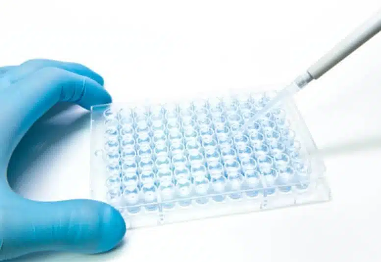
The labeled DNA is typically captured using a cell harvester on glass fiber filters, which are in turn placed in liquid scintillation counting vials for further analysis. Detection of 3H-TdR may also be conducted by autoradiography.[1]
The main advantage of the 3H-Thymidine assay is that it directly measures cell proliferation and is widely used in immunology studies. Most immune cells do not make new DNA unless they are activated, so a positive response is easily detectable against low background interference.
However, there are several disadvantages including the handling of radioactive material and relatively high cost.
Pros
- Direct measure of proliferation
- High throughput
- Clear signal-to-noise ratio
Cons
- Involves radioactive material
- Requires special instrument to detect signal
- Time-consuming and expensive
Bromodeoxyuridine Uptake
The bromodeoxyuridine (BrdU) Uptake assay follows a similar approach to measuring cell proliferation as 3H-Thymadine Incorporation assay, except BrdU becomes incorporated into replicating DNA in place of 3H-TdR and immunohistochemistry techniques are used for detection.
After BrdU is introduced to actively proliferating cells, immunodetection of BrdU using specific monoclonal antibodies allows labeling of cells in S phase of the cell cycle.[2]
After pulse-labeling cells with BrdU, mouse mAb can be used to detect BrdU incorporated into single stranded DNA.
Pros
- Direct detection of proliferation
- Non-radioactive
Cons
- Lengthy protocol
- Potential damage to cell DNA
ATP Luminescence
Adenosine triphosphate (ATP) luminescence assays measure the number of cells based on total ATP content.
ATP is a nucleotide found in cells that captures chemical energy obtained from the breakdown of food molecules and releases it to fuel other cellular processes.
Within minutes after a loss of membrane integrity, cells lose the ability to synthesize ATP, and endogenous ATPases (ATP degrading enzymes) destroy any remaining ATP, thus the levels of ATP fall precipitously.[3]
The ATP assay is based on the production of light caused by the reaction of ATP with added luciferase and luciferin. The emitted light is proportional to the level of cellular ATP in the assay, which is measured to assess the number of cells.
The glow-type signal can be recorded with a luminometer and generally has a half-life of five hours, providing a consistent signal across large batches of plates.
Pros
- Long luminescence signal (~5 hrs)
- Rapid turnaround
- No cell harvesting, centrifugation or radioactive materials required
Cons
- Requires cell lysis
- Multiple factors can cause interference with detection
CFSE Dye Reduction
CFSE (5(6)-Carboxyfluorescein diacetate N-succinimidyl ester) is a cell staining dye used for accurately tracking the proliferation of stimulated cells across multiple generations.
As CFSE diffuses into the cell, its succinimidyl group covalently binds to intracellular lysine residues, producing green fluorescence.[4] This stable linkage ensures retention of the dye within the cell, without leaching into surrounding cells.
CFSE dye provides an intense fluorescent staining which enables the tracing of eight or more generations of proliferating cells before the signal strength is diluted by intrinsic cellular auto-fluorescence.
The stable linkage and multiple generational capabilities of CFSE has led to widespread use in flow cytometry to study cell division and proliferation.
Pros
- Effective visualization of multiple generations
- Long-term signal stability
- Non-toxic, non-radioactive materials
Cons
- Toxicity may affect cell proliferation
- Low throughput
- High background fluorescence
Conclusion
Cell proliferation assays are foundational to the development of new therapies and diagnostics. Scientists today can choose among multiple techniques to measure cell proliferation that offer distinct advantages in terms of sensitivity, speed, signal stability, throughput and cost.
At Cytologics, we offer immune cell products to perform proliferation assays and conduct other immunology research. We provide HLA-typed PBMCs from healthy, normal human donors as well as immune cell subsets through negative magnetic bead isolation.
For more information or to speak with a specialist, please contact us at info@cytologicsbio.com or click the link below.
References
[1] Shevach EM. Labeling cells in microtiter plates for determination of [3H]thymidine uptake. Curr Protoc Immunol. 2001;Appendix 3:. doi:10.1002/0471142735.ima03ds21
[2] Eminaga S, Teekakirikul P, Seidman CE, Seidman JG. Detection of Cell Proliferation Markers by Immunofluorescence Staining and Microscopy Imaging in Paraffin-Embedded Tissue Sections. Curr Protoc Mol Biol. 2016;115:14.25.1-14.25.14. Published 2016 Jul 1. DOI:10.1002/cpmb.13
[3] Enkavi G, Javanainen M, Kulig W, Róg T, Vattulainen I. Multiscale Simulations of Biological Membranes: The Challenge To Understand Biological Phenomena in a Living Substance. Chemical Reviews 2019 119 (9), 5607-5774
DOI: 10.1021/acs.chemrev.8b00538
[4] Wang XQ, Duan XM, Liu LH, Fang YQ, Tan Y. Carboxyfluorescein Diacetate Succinimidyl Ester Fluorescent Dye for Cell Labeling. Acta biochimica et biophysica Sinica. 37. 379-85. Published 2005. DOI: 10.1111/j.1745-7270.2005.00051.x.