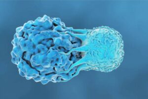
CD4+ T lymphocytes, also called helper T cells, are a type of lymphocyte that assists the stimulation of other immune cells in fighting against infection. Thus, they play a crucial part in regulating the adaptive immune response.
Immature helper T cells differentiate when the CD4+ and T cell receptor (TCR) recognize and interact with antigens presented on the cell membrane of antigen-presenting cells (APCs) by MHC II protein molecules. After activation, the immature CD4+ T cell differentiates into subtypes depending on the surrounding cytokines. During differentiation, cytokines-mediated cell signaling pathways, epigenetic alterations, and activated transcription factors are involved. [1]
CD4+ T Cell Subsets
There are six subsets of helper T cells. Each phenotype of CD4+ T lymphocytes secretes a different profile of cytokines.
- Th1. Helper T 1 cells produce IFN-γ and TNF cytokines which stimulate macrophages. They are involved in phagocyte-dependent immune reactions and cell-mediated immunity. Moreover, they encourage immunity against intracellular disease-causing agents (pathogens). [2]
- Th2. These promote B cell-mediated (humoral) immunity and produce immune responses to extracellular pathogens. They release interleukin-4, interleukin-5, and interleukin-13. [3]
- Th9. They are activated in allergic processes and helminthic parasitic infections. They produce interleukin 9. [4]
- Th17. These are pro-inflammatory CD4 + T cells present mostly at mucosal membranes. Thus, play a critical part in maintaining mucosal immunity. Th17 produces IL-17, IL-21, IL-22, IL-25, and IL-26. [5]
- Th22. They are abundantly found in the human skin. They promote the proliferation of keratinocytes and impede their differentiation; hence they play an important role in skin wound healing. Moreover, they release Interleukin-22. [6]
- Treg or T regulatory cells. They maintain peripheral self-tolerance and homeostasis by inhibiting or suppressing immune responses. Moreover, they secrete interleukin-10 and TGF-β. [7]
- Follicular Helper T cells (Tfh). They aid B cells to produce antibodies against pathogens. Follicular Helper T cells produce interleukin-21. [8]
Use of CD4+ T Cells in Research
Studying CD4+ molecular mechanisms and signaling pathways
CD4+ T cells can be a potential source to comprehend the molecular mechanisms and signaling pathways engaged in different immune responses. Understanding T cell activation and other transactivated processes can help in finding new therapeutic targets and devise effective immunotherapies against various diseases such as autoimmune illnesses. [9]
Autoimmunity and inflammatory diseases
Helper T cells are actively involved in autoimmune and inflammatory diseases such as multiple sclerosis and arthritis. T cell-directed therapies are being devised to improve these autoimmune conditions. [10][11]
Role in anti-tumor immunity
CD4+ T cells play a significant role in developing anti-tumor immunity. They mediate cytotoxic effects against tumor cells, prevent angiogenesis, and induce tumor dormancy. Thus, CD4+ T cell implications can improve immunotherapy outcomes in cancer and other hematological malignancies. [12]
Anti-viral
CD4+ T lymphocytes release different types of cytokines such as IFN-γ and TNF-α that exert anti-viral effects. These cytokines directly target the viral replication process and trigger cell death in virus-affected cells. Additionally, IFN-γ is also involved in initiating immune responses that include natural killer cells and macrophage activation that subsequently elicit anti-viral activity. [13] Additionally, CD4+ T cells obtained from convalescent patients can also be used as a potential SARS-CoV-2 treatment, providing cell-mediated immunity. Convalescent patients are those who are at the later infection stage or recovered. Similar to the concept of convalescent plasma, virus-specific CD4+ T cells are taken from the blood samples of convalescent individuals and expanded through good manufacturing protocols. Thus, later used as an anti-viral therapy. [14]
CD4+ T cells as cellular markers
CD4+ T lymphocytes are considered an important cellular marker in evaluating the stage and course of the human immunodeficiency virus (HIV) infection. Additionally, the CD4+ counts, in patients, can be used as follow-up markers to evaluate the effect of anti-retroviral treatments. [15] CD4+ T cell count is also used as an initial diagnostic test in inborn errors of immunity, characterized by immune dysregulation symptoms and infectious ailments. [16] Moreover, T cell deficiency is evaluated through numbering peripheral T cell subsets and NK cells through flowcytometry phenotyping. [17]
Diagnosis of Alzheimer’s disease
Alzheimer’s disease is difficult to diagnose in clinical setups. However, recent studies have shown that non-coding RNAs such as miRNAs can help in the diagnosis of neurodegenerative diseases like Alzheimer’s. CD4 + T cells possess many miRNAs like miRNA-let7b which is a strongly associated biomarker to Alzheimer’s disease. It interacts with other cellular genes and plays a critical role in disease pathogenesis. High levels of this miRNA are found in the cerebrospinal fluid of a patient; that is mainly due to the presence of CD4+ T cells. Thus, CD4+ T cells can be used as a potentially important biomarker for Alzheimer’s disease. [18]
Transplantation and graft rejection
Memory CD4+ T cells are involved in specific antibody-mediated graft rejection processes and transplantation failure. Recent research is using the T cell sensitization method to develop tissue or graft rejection animal models such as for renal allografts. These animal models can help explore the cellular mechanisms behind graft rejection processes and thus aid in developing therapeutic approaches for its prevention. [19]
Want to Learn More?
To learn more about this topic, check out our recent articles on T cell biology and the role of immune cells in preclinical research.
References
[1] Camara NOS, Lepique AP, Basso AS. Lymphocyte differentiation and effector functions. Journal of Immunology Research. 2012;2012. [2] Levy A, Chargari C, Cheminant M, Simon N, Bourgier C, Deutsch E. Radiation therapy and immunotherapy: implications for a combined cancer treatment. Critical reviews in oncology/hematology. 2013;85(3):278-87. [3] Nemati M, Malla N, Yadav M, Khorramdelazad H, Jafarzadeh A. Humoral and T cell–mediated immune response against trichomoniasis. Parasite immunology. 2018;40(3):e12510. [4] Sehra S, Yao W, Nguyen ET, Glosson-Byers NL, Akhtar N, Zhou B, et al. TH9 cells are required for tissue mast cell accumulation during allergic inflammation. Journal of Allergy and Clinical Immunology. 2015;136(2):433-40. e1. [5] Khader SA, Gaffen SL, Kolls JK. Th17 cells at the crossroads of innate and adaptive immunity against infectious diseases at the mucosa. Mucosal immunology. 2009;2(5):403-11. [6] Eyerich S, Eyerich K, Pennino D, Carbone T, Nasorri F, Pallotta S, et al. Th22 cells represent a distinct human T cell subset involved in epidermal immunity and remodeling. The Journal of clinical investigation. 2009;119(12):3573-85. [7] Koizumi S-i, Ishikawa H. Transcriptional regulation of differentiation and functions of effector T regulatory cells. Cells. 2019;8(8):939. [8] Cano RLE, Lopera HDE. Introduction to T and B lymphocytes. Autoimmunity: From Bench to Bedside [Internet]: El Rosario University Press; 2013. [9] Tai Y, Wang Q, Korner H, Zhang L, Wei W. Molecular mechanisms of T cells activation by dendritic cells in autoimmune diseases. Frontiers in pharmacology. 2018;9:642. [10] Skapenko A, Leipe J, Lipsky PE, Schulze-Koops H. The role of the T cell in autoimmune inflammation. Arthritis research & therapy. 2005;7(2):1-11. [11] Raphael I, Joern RR, Forsthuber TG. Memory CD4+ T cells in immunity and autoimmune diseases. Cells. 2020;9(3):531. [12] Tavakolpour S, Darvishi M. The Roles of CD4+ T-Cells in Tumor Immunity. Cancer Immunology: Springer; 2020. p. 63-90. [13] Kamphorst AO, Ahmed R. CD4 T-cell immunotherapy for chronic viral infections and cancer. Immunotherapy. 2013;5(9):975-87. [14] Cooper RS, Fraser AR, Smith L, Burgoyne P, Imlach SN, Jarvis LM, et al. Rapid GMP-Compliant Expansion of SARS-CoV-2–Specific T Cells From Convalescent Donors for Use as an Allogeneic Cell Therapy for COVID-19. Frontiers in immunology. 2020;11. [15] Mishra SK, Shrestha L, Pandit R, Khadka S, Shrestha B, Dhital S, et al. Establishment of reference range of CD4 T-lymphocyte in healthy Nepalese adults. BMC research notes. 2020;13(1):1-6. [16] Grumach AS, Goudouris ES. Inborn Errors of Immunity: how to diagnose them? Jornal de Pediatria. 2021;97:84-90. [17] Ballow M. 258 – Primary Immunodeficiency Diseases. In: Lee Goldman AIS, editor. Goldman’s Cecil Medicine (Twenty Fourth Edition) W.B. Saunders; 2012. p. 1615-22. [18] Liu Y, He X, Li Y, Wang T. Cerebrospinal fluid CD4+ T lymphocyte-derived miRNA-let-7b can enhances the diagnostic performance of Alzheimer’s disease biomarkers. Biochemical and biophysical research communications. 2018;495(1):1144-50. [19] Gorbacheva V, Fan R, Fairchild RL, Baldwin WM, Valujskikh A. Memory CD4 T cells induce antibody-mediated rejection of renal allografts. Journal of the American Society of Nephrology. 2016;27(11):3299-307.


